Open Science
We embrace the Open Science movement to make research more transparent and share data as much as possible. This means our projects are reviewed and validated by our peers and our output, whether it is software, methods or data, are made accessible to the benefit of other scientists, healthcare professionals, and patients.
Software
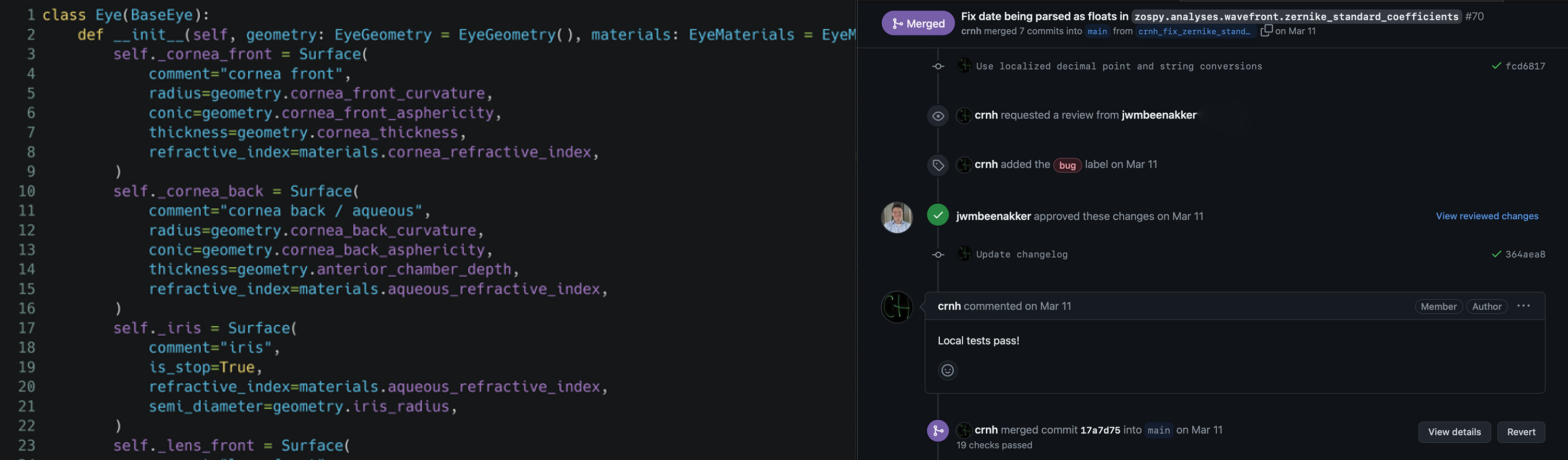
ZOSPy is our open source platform to perform optical simulations in Optic Studio and to share with other scientists through Jupyter notebooks
PAROS is a tool to calculate the magnification of fundus photographs based on the optical characteristics of the patient’s eye, as described in Correction Method for Optical Scaling of Fundoscopy Images
VFPy a python package for automated digitalization of ophthalmic visual field data


Methods
Quantitative MRI
EPG-based fit to obtain the T2water from multi-echo spin-echo MRI-scans of muscles, as described in T2 relaxation-time mapping in healthy and diseased skeletal muscle using extended phase graph algorithms
Decomposition of water and fat images from multi-echo (Dixon) MRI scans, which uses an iterative fitting method, wherein B0 field and relaxation time R2* included
Ray tracing
- Determine the peripheral retinal illumination in models of the (pseudophakic) eye, as described in Effect of anatomical differences and intraocular lens design on Negative Dysphotopsia
MRI protocols
- General ocular protocol, including DWI and PWI. ==Description, Examcard (Philips), scientific publications 1, 2, 3==
- protocol for ocular proton therapy planning [idem]
- protocol for imaging the eye lids
Data
- As many of our data, such as MRI-scans, contain potentially identifying information, they cannot be made publicly available. These data can, however, be shared with scientists who meet the criteria for access to these data and after signing of a data transfer agreement to ensure the subject’s privacy. Requests can be send to [email protected].
- Optical eye models of pseudophakic eyes with and without Negative Dysphotopsia, as described in Effect of anatomical differences and intraocular lens design on Negative Dysphotopsia
- Radiological characteristics of 26 uveal melanoma patients before and after treatment as described in MR-based follow-up after brachytherapy and proton beam therapy in uveal melanoma